|
�����p�y�[�W
���i����
���i�J�e�S��
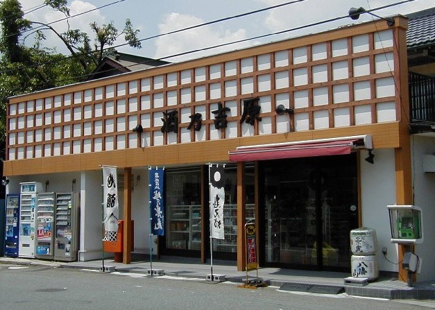
��T�g���̓X��
���Y�҂̊炪������ �S��i�������Ă���܂� �n���E�{�i�Ē��E���C�� ���g�����X ���s���Ë抁���J6-16-3
|
���̒n����w��T�g���i�g�����X�j�x�@�n���E�{�i�Ē��E���Y���C���̐��X�@�P�{�A�P����S�������v���܂��B        �^�c�҂̏Љ�
���菤����@�Ɋ�Â��\��
�l���ی�̂��߂̍s���w�j
��T�g���ւ̏ڍגn�} ���s����肨�Ԃ̏ꍇ �����Q�S�U���������ؕ��ʂɑ���A�w�����J�x�Ƃ����M�������܂��A��ڂ̐M���i�����J�w�����j���E�܂��ĉ������B���Ȃ�ɑ����čs���A�K�荕����̐M���ɂԂ����O�̓������܂���Ɠ��X���������܂��B �����w���ʂ�肨�Ԃ̏ꍇ �K�荕����𓌖����C���^�[���ʂɑ���A�w�����J�U���ځx�Ƃ����M�����E�܂��ĉ������B�E�܂��܂��ƍ���Ƀ_�C�n�c�̃V���[���[���E�E��ɑ��c�����ԗl�i�����ԏC���H��j���݂��܂��B�����ԏC���H��̗���̓��ɓ��X���������܂��B ���������C���^�[���ʂ�肨�Ԃ̏ꍇ �K�荕�������w���ʂɑ���A�w�����J�U���ځx�Ƃ����M�������܂��ĉ������B���܂��Ă����ɉE�܂��Ē����Ɠ��X���݂��܂��B�ڈ�̓_�C�n�c�̃V���[���[���ł��B �����}�c���s�s���E�����J�w����o�X�̏ꍇ �����J�w�̉��D���o�܂��āA���ɕ����Ă����܂��ƃo�X���[�^���[���������܂��B �Q�Ԃ̏�����w����w�s���x���w��������O�s���x�ɂ���艺�����B �w�������O�i�����ӂ����܂��j�x�ō~��ĉ������B�k���O���ł��B �����}�c���s�s���E�����J�w����k���̏ꍇ �w�̉��D���o�܂��āA�E�ɕ����Ă����܂��B���Ȃ�ɕ����Ė�P�D�S�������܂�B �K�荕�쓹�H�̌����_�̎�O�̓������ɋȂ���Ƃ����ł��B �k���ł��ƁA�����J�w����Q�O�����炢�����邩�Ǝv���܂��B �����}�c���s�s���E�{�O���w����̏ꍇ �s�c�o�X�w��c�c�Ə��s���x���w��c�a�@�s���x�ɂ���艺�����B ���}�o�X�̏ꍇ�ɂ́A�w����s���x�ɂ���艺�����B�w�������O�i�����ӂ����܂��j�x�ō~��ĉ������B�k���O���ł��B �����}�c���s�s���E����w����̏ꍇ ���}�o�X�w�����J�w�s���x�ɂ���艺�����B�w�������O�i�����ӂ����܂��j�x�ō~��ĉ������B�k���O���ł��B ���i�q�앐���E�V��w����̏ꍇ �s�c�o�X�w�{�O���w�s���x�ɂ���艺�����B�w�������O�i�����ӂ����܂��j�x�ō~��ĉ������B�k���P���ʂł��B
�����N�҂ւ̎�ނ̔̔��͌ł����f�肵�Ă��܂��B �����N�҈����֎~�@�ɂ��A��ނ̒ʐM�̔��ł́A���q�l�̔N��̂��L�����`���Â����Ă���܂��B �䒍���̍��u���̑����͍��ڗ��v�K���䋦�͉������B �u���̑����͍��ڗ��v�́A�u�����O�v�u���Z���v���L�ڂ���y�[�W�̉��̕��ɂ������܂��B �䋦�͂��Ē����Ȃ����q�l�ɂ́A�̔��o���܂���B�������������B Copyright(C) 2007 YOSHIHARA ,Inc. All rights reserved. �s�f�ڂ̋L���E�ʐ^�E�C���X�g�Ȃǂ̖��f���ʁE�]�ړ����ւ��܂��B�t |